Woman's puzzling decline turns out to be cobalt poisoning from hip replacement
Doctors find grey fluid and dead, metallic flesh inside poisoned woman’s hip.
A 56-year-old woman was admitted to a hospital with an array of alarming symptoms that were only getting worse. For eight weeks, she had a painful “pins and needles” feeling that started in both of her feet and then began working its way up her legs. By the time she arrived at the hospital, she was unable to feel her feet on the ground. She frequently stumbled and clutched at walls to stay up. But the tingling numbness was moving into her hands, too. Then came neurological symptoms. She told her doctors about short-term memory problems and difficulty concentrating. She was irritable and had no appetite. She was experiencing heart palpitations, too.
According to a case report this week in the New England Journal of Medicine, her doctors looked through her medical history for clues, finding nothing that immediately stood out. She had high blood pressure, a history of anxiety and depression, and hypothyroidism (an underactive thyroid). They did notice that, although she had managed the thyroid problem for more than a decade at the same dose of medication, she had been switched four weeks earlier to a stronger dose. But the dosage change didn’t immediately raise any red flags.
She also had a history of hip problems. Twenty years before, she had a hip replacement that stemmed from an injury she sustained in a car crash ten years before that. While more than 90 percent of hip replacements last at least 30 years, the woman’s started failing her after 19.
The year before her current condition, the hip prosthesis had become dislocated. At the time, doctors were able to put it back into position without surgery, but she continued to have pain and problems walking. Imaging also indicated that the lining in the hip socket was failing. So about three months before her alarming symptoms developed, she had surgery at a different medical facility to replace parts of her artificial hip joint, a surgery described as a hip “revision.”
The doctors didn’t have the medical records for the revision, but they didn’t think complications from such a surgery would explain her current condition. Of course, it’s possible the surgery could have damaged nerves, causing tingling and pain, which she was experiencing. But such damage would likely only affect nerves on one side of her body—the side with the hip replacement, which was her left side. But she had pain, tingling, and numbness on both sides. Further, nerve damage from surgery wouldn’t explain her other symptoms.
The doctors performed a thorough exam and began testing. They noted that her heart rate was elevated, a condition called tachycardia. She also had reduced sensitivity to touch in all four limbs. They considered a long list of possible causes: Vitamin B12 and copper deficiency, a rare immune disorder, chronic inflammation, and an autoimmune disease. But there were no clear leads.
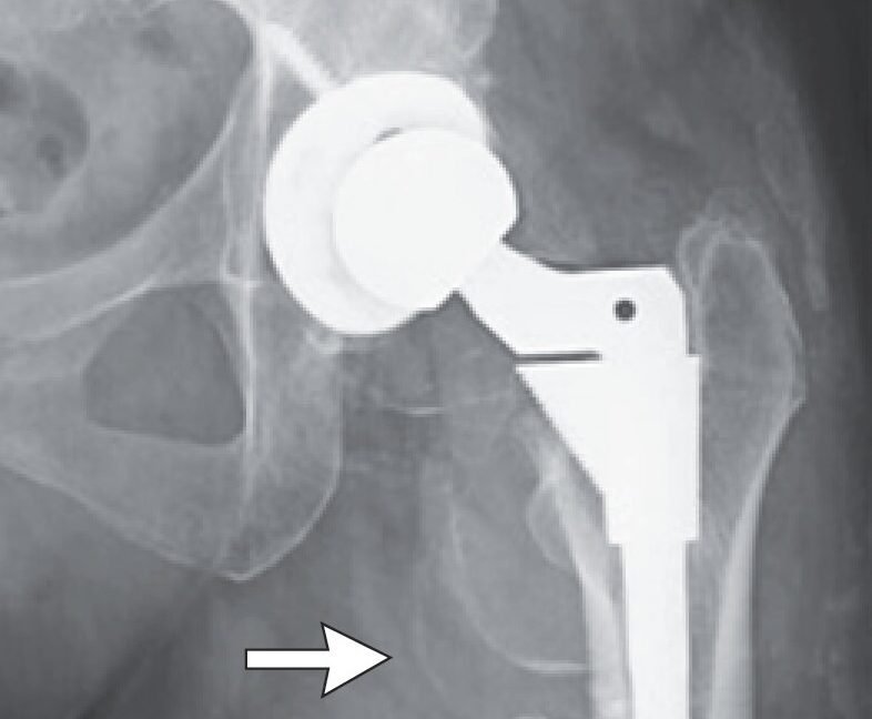
They noted that her blood work showed elevated levels of hemoglobin, an iron-containing protein in red blood cells that binds and transports oxygen. They also did an X-ray of her hip, which showed that the artificial joint was in the proper place. But it also showed deposits in the tissue around the joint.
Harrowing hip
At that point, the records from her hip revision came in. The report clarified that 20 years ago, the woman had received a titanium and ceramic hip joint. Specifically, the joint included a titanium shell (acetabular shell) that fit into the hip bone, a ceramic liner in that shell, then a ceramic ball (femoral head) on the top of a titanium stem (femoral stem) that extended into her thigh bone (femur). Over time, the ceramic liner in the acetabular shell shattered, and the ceramic femoral head began directly moving against the titanium shell.
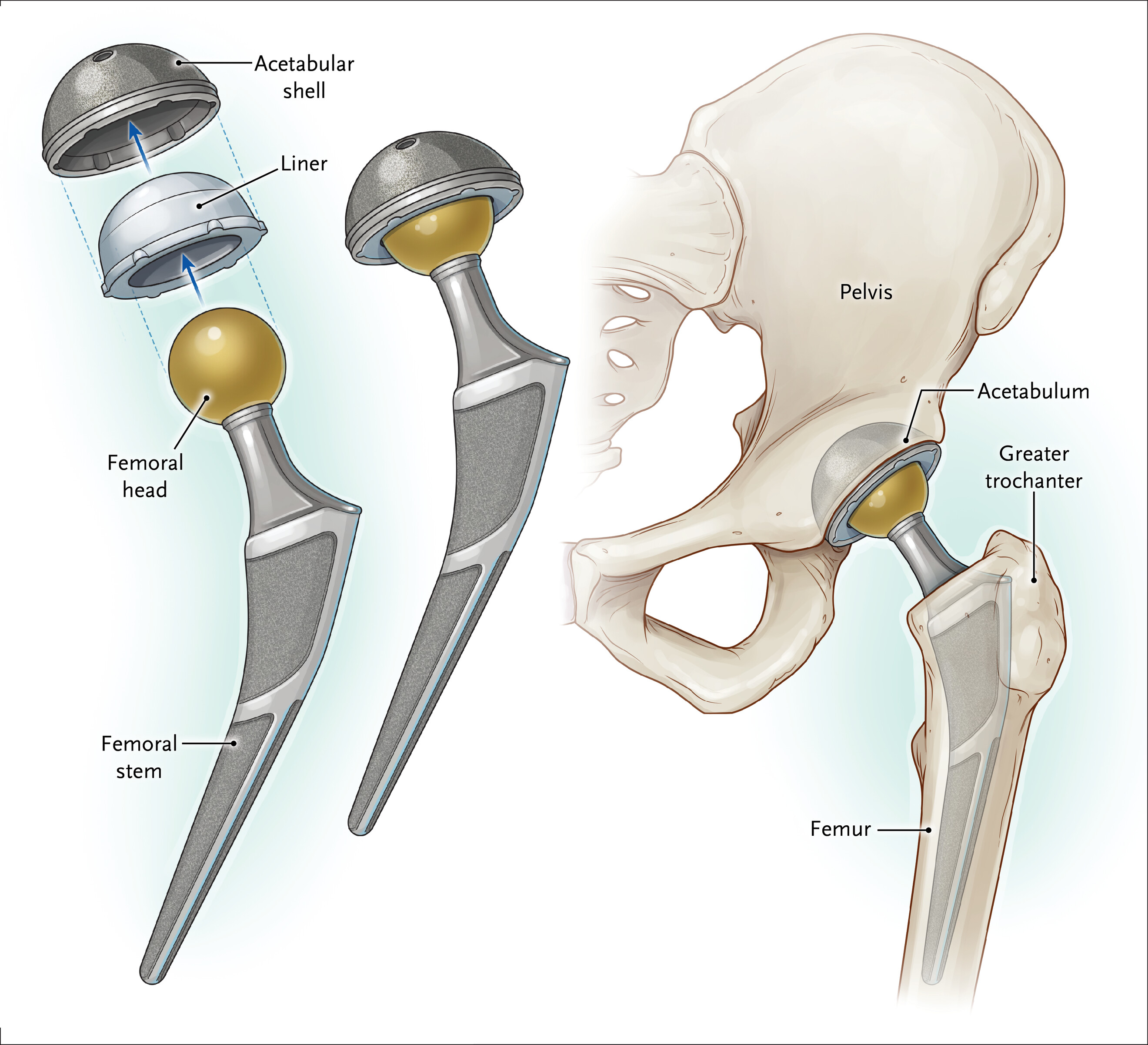
The main prosthetic components in total hip arthroplasty are shown separately, assembled, and in position within the native hip joint.
The main prosthetic components in total hip arthroplasty are shown separately, assembled, and in position within the native hip joint. Credit: New England Journal of Medicine, 2026, Bajwa et al.
During the hip revision, a surgical team replaced the destroyed ceramic liner with one made of polyethylene. They also replaced the ceramic femoral head with a cobalt–chromium alloy one. The original titanium acetabular shell and femoral stem were kept in place. The report noted that the team had to do extensive cleaning of the woman’s hip to try to clear out all the fragments of the wrecked ceramic liner that had scattered in the joint and surrounding tissue.
After seeing the report, the woman’s doctors immediately understood the problem: She had severe cobalt poisoning.
The hallmarks of cobalt poisoning fit the woman’s array of symptoms neatly. Cobalt toxicity causes nerve problems, like her pain, tingling, and numbness; cognitive impairment, like her memory and concentration problems; cardiac problems, like her tachycardia and palpitations; and thyroid dysfunction, explaining why she recently needed to have her thyroid medicine increased.
Cobalt also stabilizes a protein called hypoxia-induced factor, a transcription factor that activates specific genes to spur the production of red blood cells, usually in response to low oxygen levels. But with toxic levels of cobalt, the transcription factor is active without low oxygen levels, leading to abnormally high amounts of hemoglobin-carrying red blood cells—explaining the woman’s high hemoglobin levels.
The one thing that didn’t fit was the rapid progression and severity of her toxicity. In cases of cobalt toxicity linked to hip replacements, the symptoms usually develop over many months, not weeks, as in the woman’s case. The doctors speculated that after the revision was done, there may still have been ceramic microparticles from the previous shattered liner left in the joint. Those particles may have been grinding in the joint, causing mechanical wear on the cobalt-chromium femoral head that released cobalt into the surrounding tissue and bloodstream.
Metallic muscles
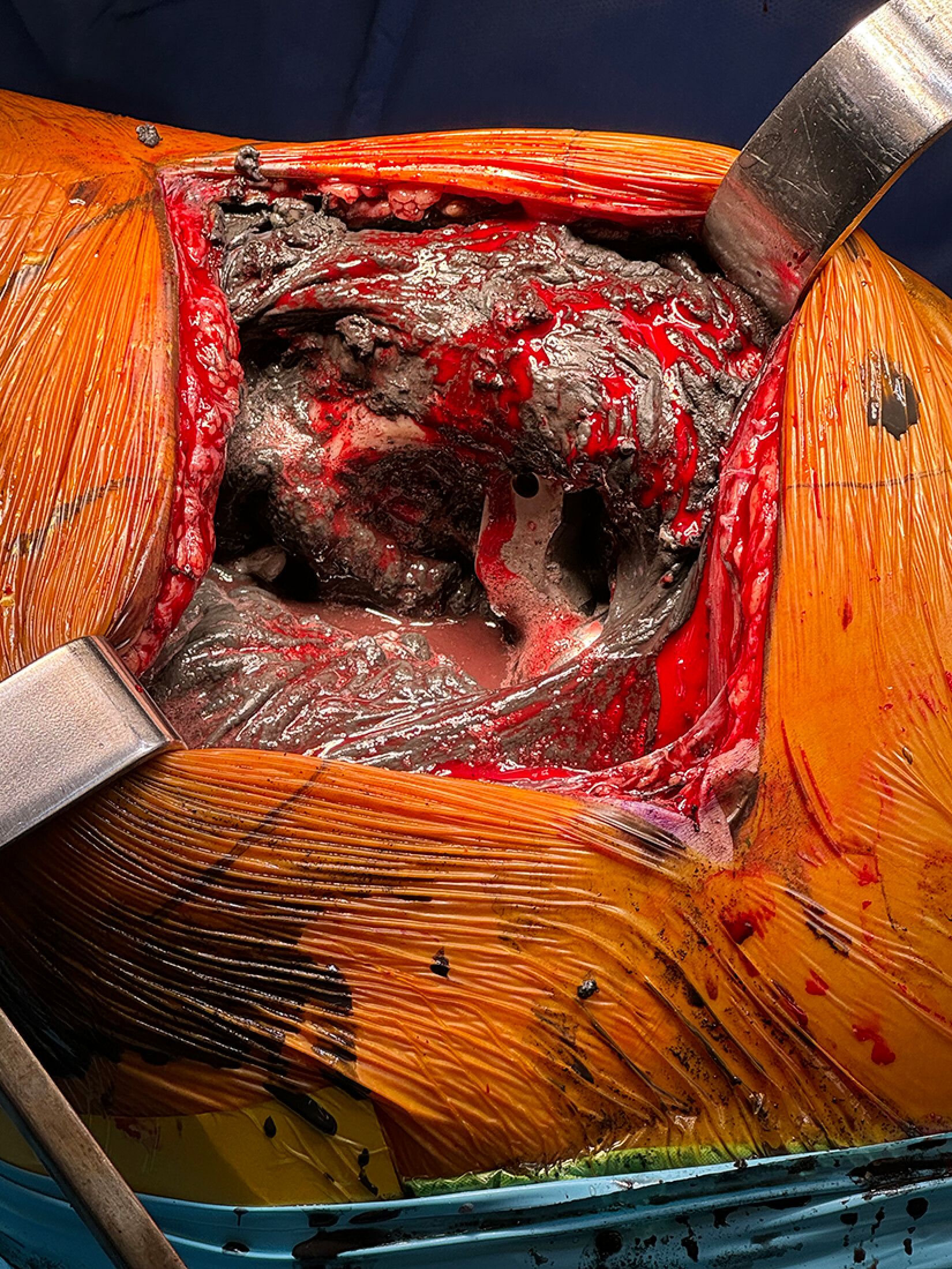
The doctors sent the woman to have a second hip revision surgery. When surgeons opened the joint, they immediately understood why her toxicity had progressed so quickly. A pool of grey, metallic fluid filled the joint while the tissues and muscles around the hip were necrotic and stained silver-gray with cobalt. (A picture of what the surgeons saw is here, but be warned that it’s graphic.)
Surgeons extensively cleaned the joint, trying to remove all of the dead, cobalt-infused tissue. They also replaced the cobalt-chromium femoral head with one made of ceramic and replaced the old polyethylene liner with a new polyethylene liner. The same day, doctors started the woman on a chelation therapy to clear the cobalt out of her body.
Three days after the surgery, lab tests came back with the level of cobalt in her blood. Before surgery, the tests found she had 592 nanograms per milliliter of cobalt in her blood. A normal value is less than 10 ng/mL. Her chromium level was 62.4 ng/mL, while a normal level is less than 0.2 ng/mL.
Her recovery was slow, non-linear, and incomplete. In the first months, her walking improved and she was able to step down her thyroid medication to her old dosage. But her nerve problems persisted. Two weeks after she was discharged from the hospital, she developed tinnitus, which also persisted. Tinnitus is a common feature of cobalt poisoning, thought to be from cobalt-mediated injury of the cochlear hair cells or the auditory nerve.
A year after her hospital stay, she reported less nerve pain, improved walking, and less frequent tinnitus.
In the case report, her doctors note that the use of cobalt–chromium alloy in hip replacements has declined “substantially” in the last 15 years. They remain in use for some purposes, though, such as certain types of hip revisions. When they cause toxicity, it’s usually due to mechanical stress over long periods and specifically involves cobalt; chromium isn’t as much of a concern. The kind of chromium used in implants is predominantly trivalent (not the hexavalent kind in environmental pollution). Chromium can damage bone near the implant, but it has relatively limited uptake by cells and isn’t linked to systemic toxicity like cobalt.
In the woman’s case, the daily grind of residual ceramic debris from her previous artificial hip significantly sped up wear-and-tear of the cobalt-chromium femoral head, which in turn sped up the release of cobalt, causing her systemic toxic illness, the doctors concluded.

Beth is Ars Technica’s Senior Health Reporter. Beth has a Ph.D. in microbiology from the University of North Carolina at Chapel Hill and attended the Science Communication program at the University of California, Santa Cruz. She specializes in covering infectious diseases, public health, and microbes.
What's Your Reaction?
 Like
0
Like
0
 Dislike
0
Dislike
0
 Love
0
Love
0
 Funny
0
Funny
0
 Wow
0
Wow
0
 Sad
0
Sad
0
 Angry
0
Angry
0


Comments (0)