

Controlling the brain with light earns a physiology Nobel
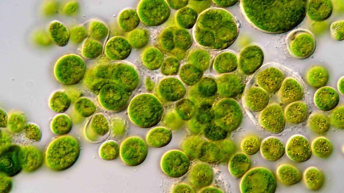
The entire field of optogenetics traces back to light-seeking algae.
Over decades of research, scientists have built up a partial picture of what specialized cells within the brain and spinal cord do. By studying how the brain develops, they could identify genes that were active in different populations of neurons and where in the brain those neurons resided. In some cases, these genes could then be used to genetically delete the neurons, allowing us to get some indication of what they might be doing, building on the information we’ve obtained from studies of brains with damaged regions.
But this approach has its limits. The brain is flexible enough to potentially adapt to the loss of some cells, and their loss early in development may alter the development of any neurons they would have normally formed connections with. It would be far more informative to activate and shut down the neurons in an otherwise intact brain.
Today’s Nobel Prize in Physiology or Medicine rewards three people—Karl Deisseroth, Peter Hegemann and Georg Nagel—who developed our ability to do precisely that. Starting from studies of single-celled algae that are attracted to light, these and many other researchers built an entire field of study that we now call optogenetics: using light to alter the behavior of nerve cells marked by the activity of individual genes.
From algae to human cells
Nerve impulses are generated by proteins called ion channels, which sit in the membrane and allow charged atoms to cross it. The nervous system uses a population of ion channels that are only active under specific circumstances, like when they sense a neurotransmitter or experience voltage changes. It’s this fine level of control that allows specialized nerve cells to send impulses only under specific circumstances, keeping the brain from descending into a haze of electrical noise.
So if we want to find out what any of the multitudes of specialized nerve cells might be doing, the easiest way is to hijack this system: force the cell to send ion-based impulses when we tell it to, and see how the animal’s behavior changes.
The Nobel Committee notes that this idea was obvious enough that people tried several methods of doing so before developing optogenetics. But it turns out “the easiest way” did not mean “easy,” and most of these methods didn’t end up widely used because they involved some combination of needing to insert multiple genes, supplying the nerve cells with some very specific chemicals, or using lasers at an intensity that physically damaged the cells.
The ultimate solution, it turned out, was lurking in a single-celled algae called Chlamydomonas. The organism’s single cell is remarkably complicated, having two flagella that help it move around, and an eye spot that detects the light it moves toward. People had been studying the organism for quite some time as a model for basic biological processes.
This is where Hegemann, then working at Berlin’s Humboldt University, entered the picture. He and his coworkers managed to hook an electrode up to a Chlamydomonas and showed that exposing it to a flash of light resulted in a very rapid influx of ions, suggesting the light was triggering an ion channel to open. As other scientists started scanning the messenger RNAs made by Chlamydomonas, Hegemann spotted a couple of genes that were similar to a light-activated ion pump found in an archaeal species.
Suspecting these might be responsible for the ion fluxes in Chlamydomonas, Hegemann used RNA interference to block their activity. This did limit the flow of ions in response to light, clearly implicating these genes in the organism’s light sensing.
At that point, Hegemann started an extended collaboration with Nagel, then at the University of Würzburg, an expert in ion fluxes. They showed that one of the two genes was selective for only letting protons (which you can think of as a hydrogen ion) into the cell, while the other would allow a broad range of positively charged ions through. They were also sensitive to somewhat different wavelengths of light. The proteins also used a chemical relative of Vitamin A to sense light, just as the receptors in our eyes do. Collectively, this class of proteins is now termed “channelrhodopsins.”
Nagel was instrumental in broadening the use of channelrhodopsins, showing that the genes that encode these proteins could work everywhere from developing frog embryos to cultured human cells. He went on to show that, when active in the neurons of a small worm called C. elegans, exposing the worms to light could cause them to alter their behavior.
Lighting up the brain
Deisseroth and his coworkers at Stanford then played key roles in developing this into a broadly useful technology. They showed that the channelrhodopsins work in neurons, and the ion fluxes they trigger are transmitted as nerve impulses. They also found more members of the channelrhodopsin family, some of which are sensitive to different wavelengths of light. A version that allows negatively charged chlorine ions into the cell was also discovered, allowing researchers to selectively shut down nerve cells.
At the same time, Deisseroth and his team worked on the engineering side of the problem, developing compact light sources and small, flexible fiber optics that allowed the system to act in the brains of animals that were free to move about and behave relatively normally.
The result has been nothing short of a revolution in our ability to understand what different populations of nerve cells are doing. If we know a gene is active in a small population of nerve cells, then we can use the mouse version of that gene to activate channelrhodopsin there and start testing how light changes the behavior of the resulting mice.
We’ve described work that relies on optogenetics a dozen times or more. And the Nobel Committee cites its use in everything from understanding how memories are stored to figuring out what signals wake mice from sleep. In one amazing set of experiments they note, a research group took an area of the brain known to be needed for maternal care and showed it contained specialized cells that performed one of a long list of functions, like triggering mothers to search for their pups, or governing physical interactions with them.
Channelrhodopsins have even been used to restore limited light sensing in a patient with a form of blindness in which their rod and cone cells die off.
One of the clearest signs of how significant this development is has been the widespread adoption of optogenetics. While the three people being honored made key contributions to its development, they have only made a relatively small contribution to its use. Instead, the studies we see that rely on optogenetics are largely done by other people who recognized its utility and refined and extended it.
The award is also very timely for the US, where the current administration has been pushing for cutbacks in fundamental research and shifting funding to its priorities in applied fields like AI and quantum computing. It’s hard to imagine a more fundamental scientific question than “how does this bit of pond scum move to ensure it gets enough light for photosynthesis?” And it’s definitely the sort of question that gets asked without any sense that the answers would ultimately have practical applications.
But science’s story, again and again, has been one where seemingly trivial research has revolutionized entire fields of study and ultimately had implications for human health and technology. The Nobel Prize Committee, in honoring a clear breakthrough, has inadvertently provided us with a reminder of that story.

John is Ars Technica's science editor. He has a Bachelor of Arts in Biochemistry from Columbia University, and a Ph.D. in Molecular and Cell Biology from the University of California, Berkeley. When physically separated from his keyboard, he tends to seek out a bicycle, or a scenic location for communing with his hiking boots.
What's Your Reaction?
 Like
0
Like
0
 Dislike
0
Dislike
0
 Love
0
Love
0
 Funny
0
Funny
0
 Wow
0
Wow
0
 Sad
0
Sad
0
 Angry
0
Angry
0












Comments (0)